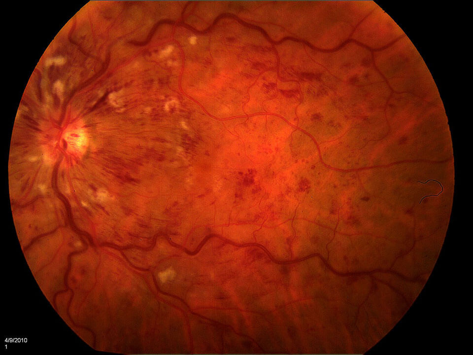
Retinal vein occlusion occurs when a vein (a vessel that drains blood from the eye) becomes blocked. A blocked vein prevents blood brought into the eye from flowing out of it effectively. The smaller blood vessels, or capillaries, that deliver oxygen and nutrients to the retina are damaged. Hemorrhages and edema in the retina are signs of this damage. A lack of oxygen delivery to the retina causes damage and decreases vision.
The loss of blood flow may also cause the eye to develop new blood vessels, which can rupture and bleed, resulting in a vitreous hemorrhage. Sometimes, these new vessels can lead to tractional retinal dislocations.
We often use a combination of intravitreal Anti-VEGF agents and laser to treat branch retinal vein occlusions. Intravitreal Anti-VEGF agents are used to treat central retinal vein occlusions. If there is a vitreous hemorrhage, vitrectomy surgery may be suggested.
There are two types of retinal vein occlusions:
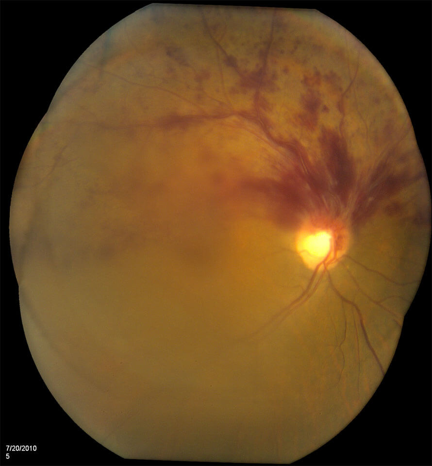
1. Branch Retinal Vein Occlusions
Before Treatment
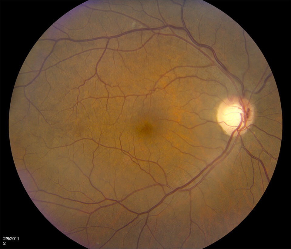
After Treatment With Anti-VEGF Agents
2. Central Retinal Vein Occlusions
- Vitreous Hemorrhage – The process and treatment are similar to those in diabetic retinopathy.
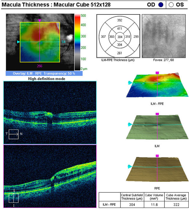
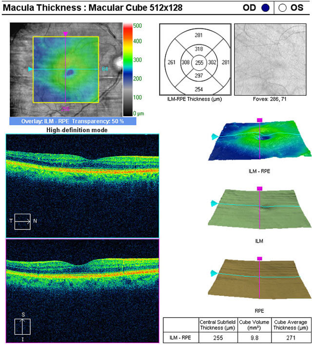
- Macular Edema
- Fluorescein Angiography – Please refer to the explanation and video at the bottom of the page.
Restore Your Vision When You Seek Retinal Vein Occlusion Treatment
Receive professional care when you contact Retina & Vitreous of Louisiana to treat your retinal vein occlusion. Call and schedule an appointment with our specialists to remedy your situation promptly.